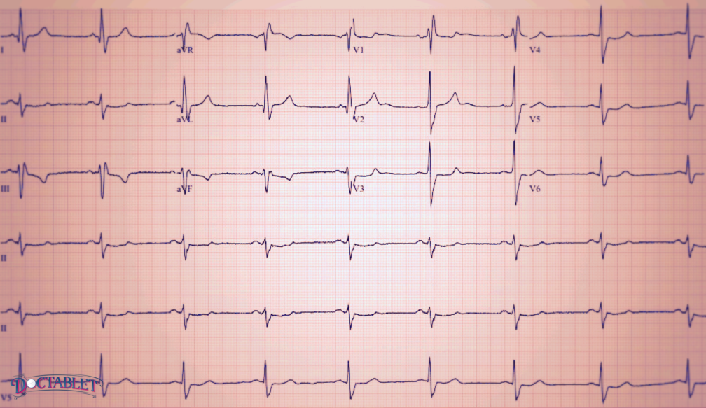
The EKG (commonly referred in German) or ECG (electrocardiogram) is an essential tool in the arsenal of cardiology and medicine. It was originally developed by Willem Einthoven in 1901, who was later awarded the Nobel Prize in 1924 for this discovery. As its name describes, the ECG machine writes down the electrical signal of the heart: electro – electrical signal, cardio – heart, gram – writing down.
The ECG is an important tool for doctors in many ways:
◉ It is easily done in the doctor’s office, taking only minutes to complete and with minimal discomfort to the patient.
◉ It is inexpensive. These days, the test can be done with any computer, with the results either printed out or stored as an image.
◉ Adds essential information to a patient’s medical visit, including the rhythm of the heart, the health of its conduction system and how fast it is beating.
These characteristics make the electrocardiogram an ideal screening test at the doctor’s office. The ECG is usually included in a patient’s yearly physical. Most people seeing a cardiologist also have one completed at least yearly, as doing so helps doctors monitor the health of the heart over time. Over the years, the science of interpreting the ECG has advanced to a significant degree. Today, doctors can accurately identify different types of arrhythmia occurring in the heart, and even pinpoint their origin within the heart’s structure with relatively good accuracy. This is helpful for people who report symptoms like palpitations and passing out. The ECG is also able to offer information about the heart’s structure, allowing doctors to get an idea of the thickness of the heart muscle; although for this purpose it’s relatively inaccurate. One of the most important functions for the ECG today is in the diagnosis of heart attacks in the emergency room, where it remains the cornerstone of that evaluation.
The different waves in an ECG represent the different parts of the heart:
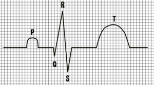
P for the atrium
QRST for the ventricle
By looking at the size and shape of these waves and the time between them, doctors can get an idea of that particular part’s structure and health. Also, specific patterns formed by these waves can be identified to further complement the analysis.
Ex: “RBBB” or right bundle branch block, Q waves